outn’about…

As part of Science Week, Alison Munro and I went on a tour of the CSIRO’s Entomology Division’s Australian National Insect Collection. Al and I share a passion for such collections.

Alison Munro, 2010 work in progress (string art!)

Al is currently a PhD candidate in the Textiles Department at the ANU School of Art. We both have a printmaking background and we met when we shared a studio during our Master of Philosophy course in the early 2000’s. Al’s current body of work is inspired by crystallography and she is researching how codes and patterns are used to construct a representation of knowledge about the natural world. As well as drawing she is learning the craft of tapestry as it relates to units of code. Since we met we have collaborated together on a few projects. My favorite was Place original face down (2004) at the ANU School of Art in 2004, where we pulled together a huge group of local artists from a wide range of disciplines; sculpture, painting, collage, printmaking and photography – all of whom use photocopy as a process in their practice. The exhibition ran concurrently with the NGA’s Print Symposium. Al and I also exhibited our work together in Super Natural (2006) in the UK. More recently we have joined with a larger group of like minded artists; Penelope Cain, Waratah Lahy, Kirstin Farrell, Ellis Hutch and Rose Montebello. We are taking our work to Level 17 Artspace at the Flinders street campus of Victoria University in October 12 this year in the exhibition Natural Digression.

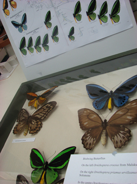
Bird wing butterflies on display at the CSIRO Entomology Division

What you immediately encounter on entering the ANIC is the intense smell of naphthalene and camphor, (I was reminded immediately of my late Grandmother) which is used as a barrier to keep pest infestations from eating everything. Beth Mantle the Collection Manager, showed us an example box devastated by pests; nothing left but the remnants and debris of what had been a collection of pretty Christmas beetles. Beginning 1928, there are now over 12 million specimens in the collection and it includes beetles, flies, bees, butterflies, moths, and other related groups such as mites, spiders, earthworms, nematodes and centipedes. We were shown display boxes of beautiful and fascinating specimens, but I would also have enjoyed sliding out the thousands of draws for a closer look.

This is a physical collection, where researchers can visit or borrow samples, but it is yet to be fully digitised, if ever. This is a problem that all museums and galleries are facing with their collections. Now that we are in the era of instantaneous web searching one just expects that an entire collection such as this should be available online. But without adequate resources, such a project will never be possible, and it could take years to record such a collection; including matching the identification tags, GPS locations, provenance etc. I imagine an entomologist would need various angles of each specimen to make it worthwhile, and consider that many insects are several times smaller than a pinhead, the tags not being much bigger. There would be various benefits of having the collection digitised, for example; expeditiously identifying exotic species in quarantine before they unleash havoc on our native environment.

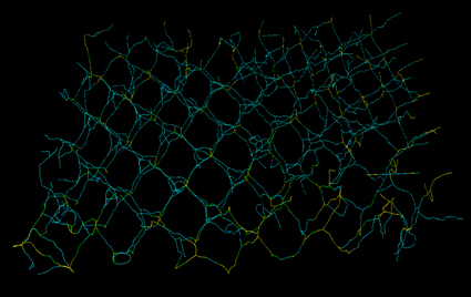
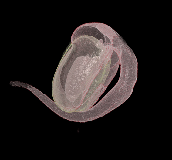
Callophris Rubi, Holger Averdunk, consultant, ANU Department of Applied Mathematics. This is one frame from a 3D animation extracted from electron tomography images of photonic crystal structures found in the wings of the butterfly Callophris Rubi.

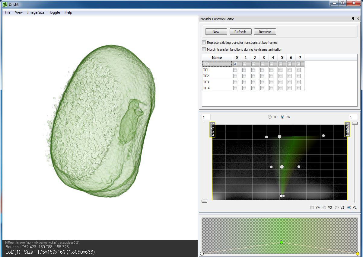
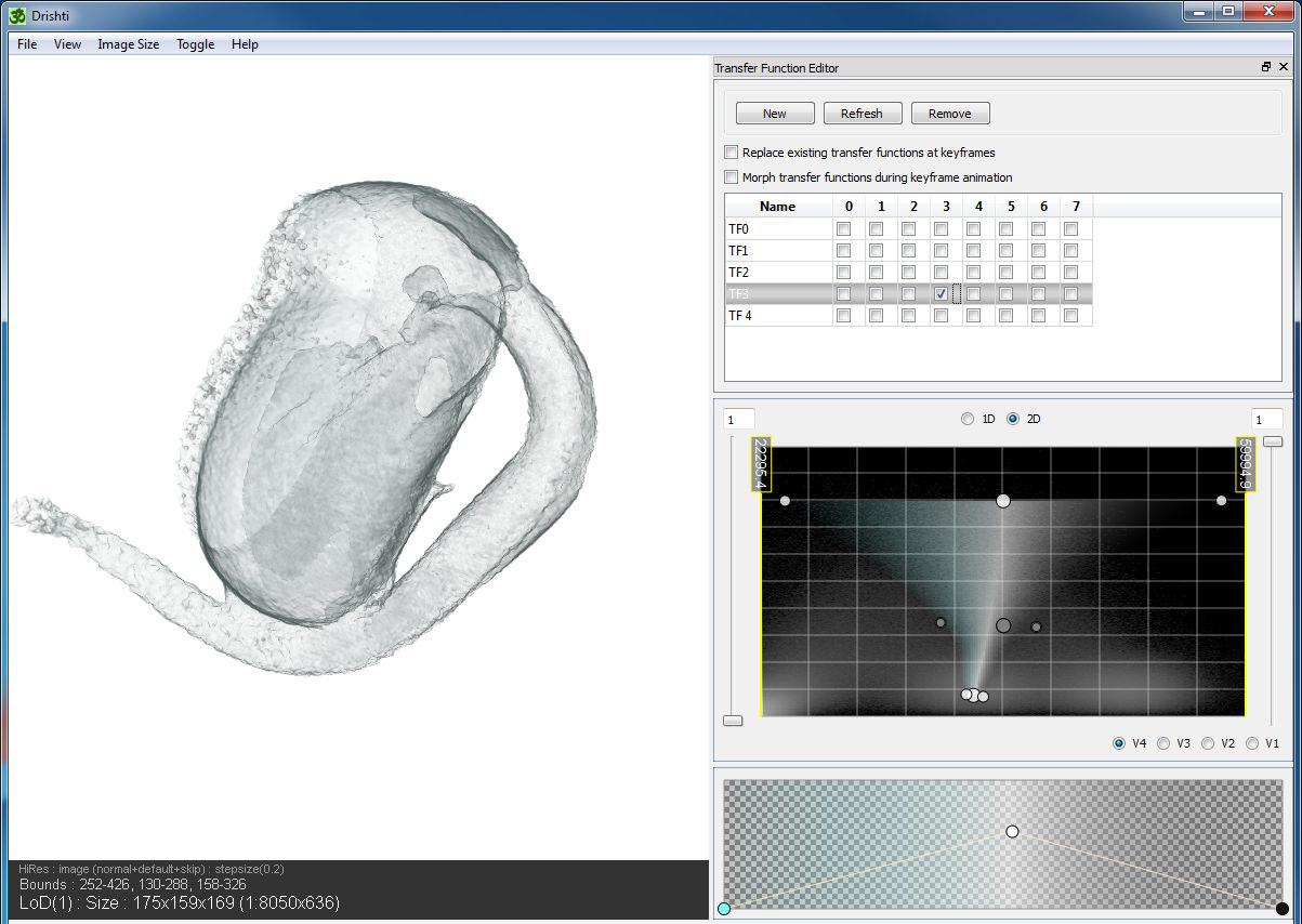
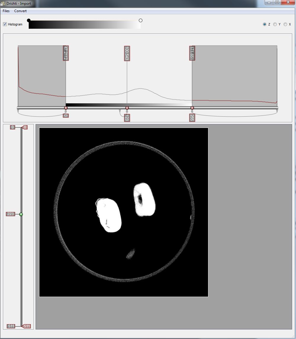
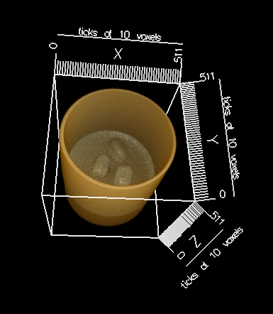
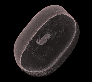
Before I decided to  looking at the sprouting beans, I had contemplated approaching the CSIRO to Micro CT a selection of specimens, but I’m not yet convinced this is the track I want to go down. But a few months ago I was in Vizlab when a number of CSIRO researchers visited to have a look at the capabilities of Drishti and XCT. Such a process would relevant to the CSIRO and many questions arose around the purpose of and possibilities of 3D XCT. A good question was asked if the Drishti software could collate and compare data – such as wing span or other biological information –  but as Ajay and Drew explained, Drishti is a volume exploration tool only. It would take long enough to photograph the ANIC let alone XCT it! There is no simple way to digitize this enormous collection. Ajay has rendered a volume of data of rhinoceros beetle, I’m not sure where it is from, but it is beautiful. The Department has also worked on electron tomography images of photonic crystal structures found in the wings of the butterfly Callophris Rubi, as it is now understood that the blue iridescence is not pigment, but the way the light reflect on the microscopic configurations.

There are a few artists who do use insects in their work. eX de Medici is of course the first to come to mind as many of her drawings and watercolour paintings are informed by ANIC having been an artist in residence in the CSIRO entomology Division since 1999. Her moth paintings symbolise the brevity and ephemeral nature of life; and while eX acknowledges natural history illustration and scientific classification, she also draws inspiration from vanitas, the 17th C Dutch tradition of memento mori painting.

Posted in excursion, research | Leave a comment

Art forum…

On Wednesday this week I presented a public lecture about my project at the ANU School of Art. I thought this would be a good opportunity to explain my journey from Printmedia to Applied Maths, my relationship with the Department, what relevance is my project to an art making practice, how the themes in my work have developed, how the technology I have access to has changed dramatically over the last fifteen years, and why it was important to receive funding from organizations such as ANAT to be able to undertake such a project.

I used this image as one example of how microscopy has influenced our imaginations as illustrated in popular culture. A still image from Them!, a 1954 American black and white science fiction film based on an original story treatment by George Worthing Yates; developed into a screenplay by Ted Sherdeman and Russell Hughes for Warner Bros. Pictures Inc.; produced by David Weisbart and directed by Gordon Douglas for the company. Starring James Whitmore, Edmund Gwenn, Joan Weldon (featured) and James Arness.

What helped consolidate my ideas for this talk is the slow process of populating my website with projects I have developed over the last 15 years. Writing this blog about my current project was extremely useful and I used some of the screen captures for Drishti that I have posted. But in reviewing essays or blurbs previously written about the work and revisiting some of the ideas I had been researching back then, I found that many of the themes in my work are recurrent; science, space travel, atomic energy, molecules, radio waves and telecommunications and scale.  I have always been challenged by wanting to express the sense of awe and wonderment in discovery, particularly in the way advancing technology was changing the pace and shape of society, without making work that ends up being whimsical.

Examples of these works are on my website www.ericaseccombe.com.au

I am often asked what the connection is between printmaking and the technology I am using now. I remember being drawn to printmaking in the early 80s because of the mechanical aspect of the image making process, the possibilities of multiples, immediacy and the democratic distribution of images. For the same reasons I was seduced by computers, for both their word processing and image making tools. It is now interesting to reflect on the recent introduction of computers to our society and how quickly we forget what it was like without them. Remember using floppy discs and having to reinsert three or four of them to save one file? It was only in 1994 that zip discs were launched with 100 MB of memory. When I worked as a free-lance designer I had an extra bag to carry around countless zip discs and my portable zip drive. Recently I was pleased to discover how I could purchase a 1 GB memory stick for less than $20.

Likewise, the computational extension of vision, which is the basis of the XCT facility, has only been made possible by the rapid improvements and capabilities of computer technology. The Department of Maths XCT Facility is supported by APAC, Australia’s largest public supercomputer centre which maintains an enormous volume of archival storage at the ANU. The storage is estimated at several petabytes, and one Pbyte is equal to one quadrillion bytes. APAC has its own air conditioning system to keep all those computers from over heating.

Another interesting example of the extension of vision through microscopy. I showed the audience this excerpt 'We are not alone', from a 2007 natural history documentary 'The invisible world.' I've linked this image to the movie on You Tube.

This talk gave me the opportunity to cover many of the questions I have about new technologies and the art making process. For example if we live in this era of accelerated visual information saturation, how it is possible that art in the 21st century can be more than just a reflection of over exposure? Or if art is directly inspired by scientific knowledge then can it reflect more than just the visual account of a body of knowledge? And what happens to art when it is made with the latest technology? What happens when that technology is superceded?

Just before the talk began Ajay came in and sat in the front row of what was becoming a very full room. I was already feeling really nervous about presenting this work; I often find it hard to talk about my own work in context of what the Department is doing and I worry that what I am doing is not particularly interesting in comparison. Or that I’ll say something totally stupid, or someone will ask me a really hard question. And I fret that my work is boring. However, my fears were unnecessary, it went well and it was good for me to put my practice in perspective.

Most importantly Ajay really liked my animation of the clipping planes. He’s challenged me to make a similar demonstration of using the cylindrical crop tool.

And thank you to Waratah Lahy, Gordon Bull and Anne Brennan for the opportunity to talk a the School of Art!

Posted in presentation, research | Leave a comment

counting beans…

There was a news article circulating this week about Ron Sveden of Massachusetts who thought he had lung cancer but luckily for him the growth turned out not to be a tumour but a pea sprouting; proving that those hardy crop seeds will grow in the most extreme conditions!

Thanks to the team, I now have the first image and the last image of the seed sprouting sequence. So here you can see what the two end captures look like if they their transfer functions are overlayed.

The first and last 3D volumes of the seed spouting sequence rendered in Drishti. The third image on the right is the two volumes overlaid as transfer functions.

Tim selected and set the base volume so that each new frame can be positioned in a template. Later I watched Ajay manipulate the transfer functions in order to isolate the beans from the bed of agar. He made some suggestions and showed me how he would do it by using the crop tools in combination with the transfer function. He does it expertly and I follow his methods on my own from scratch, taking me a full day to correctly capture one bean compared to Ajay’s 15 minutes.

I met two Palaeontologists in Vizlab this week, Dr Gavin Young and Alice Clement who are working with Tim on the XCT capturing the most amazing volume data of a lung fish, comparing aspects of the nervous system between live species and ancient fossils. They had come in for a lesson on Drishti with Tim, and we shared a moment commiserating on our bumbling efforts in comparison to the masterly skills of Ajay. One of the difficulties experienced by infrequent users (or even frequent users) is that Ajay constantly improves Drishti’s tools, renovating the user facilities.  So is important to be able to understand what the updates are and how they work for the data at hand. I have learnt to just persevere while referring to Ajay’s handy online instructions; then it is possible to succeed no matter how clumsily. Drishti is freeware and is now available to down load to PC or Mac so researchers are not limited to working at Vizlab, but there I have the added advantage being a stone’s throw away from Ajay.

a bean sprout - imaged rendered in Drishti.

I think it is amazing that we are looking inside a tiny bean. We predict that the difficulty we will encounter with the final bean sequence, when we proceed with animating the full dynamic process of sprouting, is that every volume in the sequence has a unique transfer function. For the moment it is not such an issue with only a few volumes but it will begin to cause difficulties with more than 5. We may have to rethink how they are linked together. However for the moment its giving me an excellent opportunity to hone my Drishti skills. To get a cleaner image I isolate one bean and clean it up as much as possible so the interior is visible through the fine outer skin. I have rendered an image (above) of the bean and its sprout overlayed, the two volumes differentiated by the colour yellow and pink. And below are two screen shots to compare the transfer functions of the first and last volumes before they merge.

screen shot of first bean in Drishti illustrating the transfer function as a histogram

If you take note of the white shadows in the histogram you can see how I’ve selected the space in between. This is the thin corridor of material between the agar jelly and the bean’s starchy interior. It allows me to examine the transparent outer shell while viewing internal cavities.

A screen shot capturing the end sprout volume in Drishti.

In this screen shot you can observe the comparative transfer function. It is different because the fragile sprout is not contrasted enough with the agar. You can see how the end of the sprout is a bit messy from being embedded with the base.

Posted in project | Leave a comment

sowing the first seed…

Finally the mungs make their debut on the XCT. They were ready, soaking over night; two mungs and one little radish for good luck. Amazingly they all sprout and don’t die halfway through the sequence – nine hours of 64 captures in 360°. Will the data be any good though? The acquisition of data is now in the hands of Dr Andrew Kingston to process the information for each shot. One data set takes a couple of hours to compile manually and the process is about to be automated in order to get through the enormous amount of work that the facility is generating.

3D tomograph of beans in Drishti import.

But now for a sneak preview. This is what the volume looks when it is first imported into Drishti to create a subvolume. The import main window is divided into two– a histogram window and an image window. The histogram window is used for display of 1D histogram of the loaded image. Having to understand 3D data as a histogram has been a mind bending experience but I’ve managed to plod along. The image window displays the currently selected slice of volume, like any tomographic image, but you can run through from top to bottom by using the left slider. Sizing the parameters of the volume cuts out most of the junk around the image, reducing the size of the volume.

First volume of beans in Drishti

As soon as a subvolume is saved I can drag it across to render. This is the full dataset, the beans in their container on a bed of agar jelly. In Drishti one would normally reduce the volume first by cropping in the low-res mode, but I’ve prepared an animation demonstrating the use of clipping planes to reduce the volume in high res. The process of cropping and clipping is usual for any 3D data set. It is about refining the image and working out which bit of the data is the important information. For instance, if you were examining closely the lung of a rat, you wouldn’t necessarily need its tail. This is what makes Drishti important as an volume exploration tool as it has the function to identify and select the data according to its material density ie; skin, flesh, fat, tissue, muscle, veins, nerves, bone, marrow…

the first bean

So what we have come to realise by looking at this first datset is that the agar jelly is too close in density to the starch based bean. This makes it difficult to use the transfer function to isolate the bean. It was easier with the plastic objects, the data was so straight forward. All I had to do was crop the stems they were attached to. The bean is different, a bit messy, no clean lines and so the object will not be whole in the way that I have imagined it. Drew in Vizlab explains that this is typical of working with living or organic matter, it is never how you want it to be and is always messy. Bones, rock and plastic are easier to render. But for now, this is all I have so I practice stripping of all the spongy muck down to the bare, transparent skin. There is a delicate shape to the bean, and a glimmer inside of the tiny shoot beginning to develop. I like it. I can’t wait to play with the rest of the sequence.

Tim suggests that in the meantime we work out another more contrasting base that will give the bean sustenance, but will not be too dense. I do a web search on such possible materials hoping that I’ll stumble across some new plant growing technology. I spend a bit of time reading about hydroponic systems and pages posted by bromeliad and orchid fanatics. Tim is an expert on all of this anyway as he has maintained his childhood passion for carnivorous plants; two enormous baskets of twisting vines hang from the ceiling of his office resplendent with snappy blooms. Tim decides to foam up the agar so it sets with air. Adding air to agar jelly makes me think of whisks, this makes me think of marshmallow which leads to Pavalova. Its not such a silly idea apparently. Perhaps whipped egg white with agar might do the trick, as long as it retains a marshmallow texture, the tiny bubbles of eggwhite might contrast with the starchy bean.

whisking egg whites in the wet lab

preparing a contrasting base for beans

I xxx
I am fond of making the dessert Pavalova and it has been an interest between my friend Rowanne and I for some time now; we have even considered blogging (Pav lovs blog) our efforts for every time we create one the result is unexpected – hit & miss. So for me whipping up eggwhite in the wet lab is seriously amusing. Lab coat, eye protection, glass test tube etc.  I get to measure out the agar with nice little measuring spoons but I bring in my own bar mix. There are magnetic mixers in the lab but it’s easier with the electric whisk. I make a few bases in Petri dishes and put them in the fridge for later. I put a few more beans on the soak.

Posted in project | Leave a comment

minute bodies…

Toward the prosecution of this method in Physical Inquiries, I have here and there gleaned up an handful of Observations, in the collection of most of which I made use of Microscopes, and some other Glasses and Instruments that improve the sense; which way I have herein taken, not that there are not multitudes of useful and pleasant Observables, yet uncollected, obvious enough without the helps of Art, but only to promote the use of Mechanical helps for the Senses, both in the surveying the already visible World, and for the discovery of many others hitherto unknown, and to make us, with the great Conqueror, to be affected that we have not yet overcome one World when there are so many others to be discovered, every considerable improvement of Telescopes or Microscopes producing new Worlds and Terra-Incognita’s to our view.  [Robert Hooke 1665, Micrographia: Some Physiological Descriptions of Minute Bodies Made by Magnifying Glasses with Observations and Inquiries Thereupon.]

Scheme 1, from Micrographia, an illustration of Hooke's microscope

Micrgraphia, by the English physicist Robert Hooke (1635-1703), is the first book on observations of the natural world through microscopic and telescopic lenses. While studying the heavens above, Hooke also looked more closely at things on Earth. He was the first person to use the word “cell” to identify microscopic structures which he discovered by looking at a thin slice of cork through a magnifying glass of his own construction. As he explains in his chapter “Observ. XVIII. Of the Schematisme or Texture of Cork, and of the Cells and Pores of some other such frothy Bodies.”;

I with the same sharp Penknife, cut off from the former smooth surface an exceeding thin piece of it, and placing it on a black object Plate, because it was it self a white body, and casting the light on it with a deep plano-convex Glass, I could exceeding plainly perceive it to be all perforated and porous, much like a Honey-comb, but that the pores of it were not regular; yet it was not unlike a Honey-comb in these particulars. [Hooke, Micrograhia]

Picure 23 - Observ. XXXVII. Of the Feet of Flies, and several other Insects.

Scheme 23 (fig 23) from Micrographia, Observ. XXXVII. Of the Feet of Flies, and several other Insects.

Hooke was observing a world yet unrevealed, and 346 years later with more capable technology at hand, we are still driven to ascertain the truth about things, known and unknown, down though the layers of cellular and subcellular structures.

Discussing Hooke, Vince Craig (Dept Head App Maths), told me that the action of the cantilever in Atomic Force Microcopy works on the principle of Hooke’s Law of Elasticity. The cantilever, (between 50 – 200 micrometres in length) is the common sensor of the force interaction in atomic force microscopy, and it is the movement of the single point at the end of the cantilever (like a needle on a record player) that studies the topography of surfaces at micro- and nano- scale.

In Hooke’s early investigations and theories in mechanics and physics he discovered the principle behind the action of a spring and developed a simple equation expressing the force generated within a compressed spring in relation to the stress or deformation of material. Hooke invented the balance spring consequently enabling the mechanical workings of clocks to keep regular time. But the curious fact is, that while Hooke examined magnification, it is only now with the invention of more sophisticated technology that the mechanism based on his law of elasticity is utilised for the purpose of microscopy in the AFM, without the glass lens.

There is an AFM at the back of the wet lab and I recently sat in on an instructional session with Tim Senden as he explained how to use the AFM to a group of researchers who needed to use this equipment independently for their own purposes… just adjust that part there without snapping it off, make sure your sample is clean, and there, see how easy it is, etc. Perhaps I missed several of the more technical tips, but it was interesting to learn the basic principles of how an AFM works.

Vince’s images from the AFM are on my website. I finally uploaded the exhibition RAW from Science Week in 2007, that brought together the work of 17 researchers from the ANU Department of Applied Maths, and took it to the School of Art in Photomedia. RAW has some excellent examples of scientific research using contemporary technology and computer visualisation; including images from the AFM and the XCT, mathematical equations, along with technical diagrams and photographs of scientific instruments which they custom build within the department.

I am fascinated by the ideas of magnification of the microscopic and I’ve always been in awe or fear of radiation or atomic power; perhaps because of my age, nuclear war was a hot topic when I was a kid and the idea of it used to terrify me. Recently when I started posting some of my previous projects on my website,  I realised how much of my work is influenced by the above concepts. An excellent example of this is an experimental piece I made in 2001 where I enlarged a tiny image of a plastic praying mantis into a seven by three meter long work entitled luvamuncher. There are many connections with my past work and the project I am undertaking now.

Posted in research | Leave a comment

back in the lab…

How do you make a mung bean remain still and at the same time keep it from drying out? The difficulties sprouting a mung bean for 3D tomography is that they flip over when they turn their sprout down. Mung beans are a hardy variety and they move quickly once their sprout gets going. Because of the rotating bed of the XCT, the object has to be secured on the same axis to remain in focus all 360 degrees. To achieve the clearest image, the base material also needs to contrast from the bean. Water is not an option as surprisingly it is too dense, so Tim proposes to try first a bed of agar jelly. We go to the wet lab to mix it up.

There is something crazy about the wet lab in Applied Maths; not the extreme sterile environment you would expect,  but it has a fantastic experimental atmosphere of tinkering around among countless glass vials. I made a documentation of it in 2006 to try and capture the essence of it and it certainly hasn’t changed much since then.

fluoroscein c=o, 2006,

Recently I viewed the work of German artist, John Bock’s film Fischgrätenmelkstand kippt ins Höhlengleichnis Refugium, (2008 video) at the MCAs, The Beauty of distance: songs of survival in a precarious age, at the 17th Biennale of Sydney. (I am yet to find an official English translation of this title, but my step son fed it into a translation tool with truly hilarious results.)  This work reminded me of the wet lab possibly because I’m viewing it from an artist’s or outsider’s point of view; I’m quite sure the Department does not view their laboratory in quite the same way!

Bock’s absurd and humorous film features an 18th century wigged and powdered inventor and his beautiful but trapped accomplice. Highlighting the complexity of language, narrative, history, performance and sculpture in the pursuit of scientific endeavor, the film is a satire evoking Descarte’s treatise Discourse on the Method of Rightly Conducting One’s Reason and of Seeking Truth in the Sciences (1637). I’ve found the most current review with images of Bock’s work  at de de ce blog.

In terms of my project I found another work at the MCA, Victoria, (2008 video) very relevant and moving. By the Finnish artist Salla Tykkä, Victoria is a ten-minute timelapse on the life cycle of a giant lily as it unfurls its petals in the dark, changing colour from pure white to a red hue as the morning light arrives. This species has very large leaves that can span up to three meters in diameter and the flowers can open up to forty centimeters wide on seven to eight meter stalks. Coincidently this summer I had visited the Adelaide Botanic Gardens and discovered for the first time the Amazon Waterlily Pavilion which is used to care for and propogate the Victorian Amazonica which was introduced into the gardens in 1868. The genus was named after Queen Victoria by John Lindley in October 1837, when the first known specimen was returned from a exploration of the South America’s Amazon (formerly British Guiana). Sadly the lily was not in flower during my visit, however, in the late 1800s the very first bloom of a Victorian Amazonica caused an enormous sensation in Adelaide. So for me, Tykkä’s video incorporated so eloquently all that has passed in terms of the history of observation of the natural world, including the beauty and the wonder.

I quite like this review, The Beauty of Distance in Kant’s aesthetics by Daniel Wilson, especially in consideration of Salla Tykkä’s companion film Airs Above the Ground, which was projected next to Victoria. Both films examine the colour white and the human fascination with beauty and the desire to capture and control nature.

Anyway, Tim and I mix up the agar and put it in the fridge to wait over night. I put a few beans on to soak to be ready; we have joined the queue. Getting on the XCT is the tricky part, the facility is in great demand. Meanwhile I spend time observing and learning the process of Micro 3D X-ray.

Dr Michael Turner preparing samples for the XCT

Dr Michael Turner is in there mostly and he tolerates my presence. We discovered we are in fact distant cousins, so he is obliged to put up with me, but Michael is really very inclusive and I’ve come to understand more about the process by sitting next to him and just watching. There is a lot of adjusting. When I began my initial residency the XCT was the first and only machine in the facility. Since then the team has advance the program so they have a bit more external control – before they had to walk in and out and re-program every time. Now they have included a new XCT with a helical base, this means that they can capture more information and larger sections of data. For example, a length of bone or rock, rather than a tiny sample.  But this second XCT is not yet fully operational. They are also working on a third room and it already has its lead lined walls installed and painted.

Posted in project | Leave a comment

mung bean…

I felt very anxious about starting again in the Department of Applied Maths. Last time it seemed so easy; those plastic objects were perfect. Back then I was more worried how I was going to make work with 3D data, that was the challenge. But what to X-ray now? The team are looking at dynamic systems and that means rock and bone. I was really worried that Tim might think my sprouting seed idea rather simplistic. The Department meets for morning coffee and welcome me back. Naturally I am asked what I’m planning to do this time round. I felt so self-conscious I waited for everyone to go back to work before I proposed the idea to Tim; will 3D X-rays of sprouting seeds work? I don’t know why I was worried. Tim had of course thought about it before and can’t wait to give it a go.

My original idea was to X-ray Australian native seeds as they germinate – as they have curious structures I thought they would be visually interesting – but to do this I would have to become an expert on how they sprout. For the moment we need something immediate. Mung beans are the obvious choice; they are so willing to grow they will sprout happily in space. Infact, when I look them up there are a some sites proffering school experiments on how to sprout mung beans rotating on record tables to simulate anti-gravity. Without gravity, where do mungs direct their sprouts? In space, do they know what is up or down?

So first things first. I go to Dickson to the health food shop in search of seeds. A nice girl in a loose cotton shift helps me choose a box of organic bio-snacky mung beans. She looks pleased and makes a comment about my healthy choice. I’m not sure if I can tell her and I feel strangely guilty when I hand over the cash; its not like I’m about to torture mice…

But how do mung beans grow? There are excellent sources available for timelapse photography and this example was filmed by the science technicians of Reigate college in the UK over aprox 10 days.

Mung bean germination – HD timelaspse

I start growing a few in a bowl on the kitchen sink at home. In theory I know what the seeds do but now I have to be a bit more observant. They expand considerably in the first 12 hours of soaking. I rinse the water and keep them moist. I have to be out all day so when I come home they have expanded some more. The next morning they have split their green jackets and I can see a tiny protrusion. hmm, I have to say it does look a little bit like a penis. It only takes a few days and all of the mung beans have three to four centimetre sprouts. I wonder what they will look like X-rayed.

Posted in project | Leave a comment

from the beginning…

I am a practicing visual artist who lives and works in Canberra. My background is in printmaking and my work is informed by the way scientific technology has influenced visual media and contemporary art. You can view more of my work at www.ericaseccombe.com.au

My application to Synapse Residency Program is based on the strength of the collaborative relationship I have already established with the ANU Department of Applied Mathematics in the School of Physical Engineering and Physics.

3D microcomputed tomograph X-ray volume of a plastic Nudibranch in Drishti

I initially approached the Department in 2005 and in 2006 I commenced a 3-month artistic residency supported by an artsACT project grant. At the time my residency coincided with the preliminary stages of the Departments XCT Facility and I was fortunate to have the support of Associate Professor Tim Senden who facilitated the XCT scanning of a selection of my collection of miniature plastic animals. To visualise and animate these datasets I was required to learn how to independently use Drishti: a volume exploration and presentation tool developed by Dr Ajay Limaye. At the time Drishti was in its formative state I was the primary user and tested various capacities with each new upgrade; an intensely challenging process. The result of this association can be seen in my subsequent works Nanoplastica (2008) and Ocular (2006). (image Slug, part of the Nanoplastica series, 2008)

Nanoplastica installation at Canberra Contemporary Art Space, 23 May - 5 July 2008, site-dimensions; CCAS main gallery 20390 cm wide x 8750 cm depth, with a ceiling height of 4450 cm from floor to the beginning of the roof beams.

Nanoplastica was exhibited at Canberra Contemporary Arts space (2008) and then later projected on the BIG SCREEN at Federation Square, Melbourne, (2009). The first edition of Nanoplastica was acquired by Canberra Musuem and Gallery in 2008. Ocular (2006) the first work in this series of 3D animations, was exhibited in the exhibition Super Natural, at the Alsager Art Centre in Manchester University (2006). Nanoplastica is also included in the exhibition Linear Progressions as part of the National Science week beginning 14 August 2010, screening at QUT Creative Industries Precinct Parer Place, Kelvin Grove Urban Village screens, selected Screens at Gardens Point & Federation Square.

On the strength of these works and investigation, I returned this year to the Department of Applied Mathematics as a Visiting Fellow supported by a 2010 artsACT project grant. And now the Synapse residency program has enabled me to continue this project in depth at a collaborative level.

The Department of Applied Mathematics is mix of research physicists, chemists and mathematicians with broad interests in the form and function of matter. They are recognised for groundbreaking research into processes involving complex materials and networks. Already their results are challenging theoretical knowledge of the structure of matter, including the established understanding of evolution. The Departments technology and software has greatly extended their experimental capabilities, causing scientists from a wide range of disciplines to re-evaluate how they now approach their research. The facility offers the highest fidelity computer tomography possible today. These XCT instruments were designed and built in-house and they scan objects in 3D datasets, (not in slices). The Facility is supported by APAC, Australia’s largest public supercomputer centre which maintains an enormous volume (several Pbyte) of archival storage at the ANU. Through the XCT the Department has attracted a wide range of business partners and is now an advisor and supplier of this equipment to similar research and industry facilities throughout the world.

My proposal to Synapse is to collaborate with the Department of Applied Mathematics’ Dynamic XCT project. A team is currently researching the visual possibilities of computer tomography in microscopic kinetic systems; such as limestone rock dissolving or water wicking through fibre. Scanning of static objects is now considered routine while research into new methods for studying dynamically changing systems in 3D on the microscopic scale is just beginning. My collaboration will focus on two aspects of their data.

Firstly, the understanding of the complex interrelationship of mechanical tensors in 3D. The 3D fabric of a rock or bone encodes the mechanical properties of the object.  The complex interplay between structure, form and material composition can only be solved using a super-computer. For a material under stress each point in space can be described with 9 different components, or tensors. Understanding how each tensor correlates to a particular mechanical property could greatly improve interpretation. Turning 8 billion volumetric data points, each point possessing 9 properties, into a meaningful image involves the use of animation, and a deep understanding of colour and light.

The second aspect of their new experimental capability is the acquisition of 3D in cinematographic mode.  The ability to study the time evolution of 3D systems such as moving sand grains, fluids flowing in rocks or seeds geminating is only marginally feasible at present.  This group is soon to patent a novel method for acquiring 3D datasets in very short times. The visual density of grains moving in a sand pack, for example, is far too great to observe unless the key parameters are singled out.  These might be, in this case, the portrayal of the rotational axis with time. Systems in which 150,000 grains are tracked look dense and intractable.

With this project the Department is aiming to balance visual density with information content. Scientists are used to graphing data, however in 3D, and in particular multi-dimensional data, the portrayal of dense datasets is not a simple task; the inter-relatedness of properties and structures maybe complex and non-linear. The team recognises that perception is guided by a visual sense, and the group is developing a visualisation toolbox to enable this next leap forward.

Much of the Departments research is driven by international oil and gas company ventures, which in turn validates their investment in the XCT facility. As part of my collaboration I am also proposing to contribute to the Dynamic XCT project by scanning the germination process of seeds for my own artistic purposes. In 2006 my plastic objects became perfect sample datasets for assessing the various associated visual systems and demonstrating data visualization across disciplines. My new proposal will be a good trial product for the team because organic matter has very little density in comparison to rock; the scanning time will be reduced, datasets more readily understood, and the dynamic sequences theoretically predicted.

Posted in research | Tagged | Leave a comment